90 results
- Books
- Online
A most certaine and true relation of a strange monster or serpent found in the left ventricle of the heart of Iohn Pennant, Gentleman, of the age of 21. yeares. By Edward May Doctor of Philosophy and Physick, and professor elect of them, in the colledge of the academy of noble-men, called the Musæum Minervæ: physitian also extraordinary unto her most Sacred Majesty, Queene of great Brittany, &c.
May, EdwardDate: MDCXXXIX. [1639]- Pictures
Congestive heart failure in a 70-year old man with digitalis, dyspnoea and chronic abdominal distension: surgical specimens of (a) embolism in superior mesenteric artery, (b) intra-mural thrombus in left ventricle and (c) diagram of gangrenous small intestine. Watercolour by Barbara E. Nicholson, 1951.
Nicholson, BarbaraDate: 1951Reference: 34211iPart of: Barbara Nicholson medical illustration collection.- Pictures
Widespread cancer in a 50-year old man with fatal bronchopneumonia: detail sections of (a) left kidney, with fungus like growths, (b) left heart showing metastases nodules, and (c) right heart encompassed in yellow tumours. Watercolour by Barbara E. Nicholson, 1951.
Nicholson, BarbaraDate: 1951Reference: 34318iPart of: Barbara Nicholson medical illustration collection.- Books
The spread of the excitatory process in the toad's ventricle : preliminary communication / by Thomas Lewis.
Lewis, Thomas, Sir, 1881-1945.Date: [1915]- Videos
- Online
Cardiac irregularities.
Wiggers, Carl J. (Carl John), 1883-1963.Date: 1929- Books
Lidocaine in the treatment of ventricular arrhythmias : proceedings of a symposium held in Edinburgh in September 1970 / edited by D.B. Scott and D.G. Julian.
Date: 1971
- Digital Images
- Online
Medullary cancerous tumour of the endocardium of the heart
Delamotte, William Alfred
- Digital Images
- Online
Heart with aortic valves ulcerated and disorganized
Godart, Thomas
- Books
- Online
Case of a small aneurysm of the first part of the arch of the aorta, opening into the pulmonary artery and conus arteriosus of the right ventricle : with remarks on the general subject / by Sir William T. Gairdner, M.D., K.C.B., F.R.S., Physician to the Western Infirmary, Glasgow.
Gairdner, W. T. (William Tennant), Sir, 1824-1907.Date: [1899]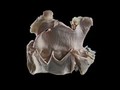
- Digital Images
- Online
Aortic valvular endocarditis, horse. This equine (horse) aorta has been dissected at the level of the valve, separating the artery from the left ventricle (bottom chamber). Nodules are present which have formed as a consequence of endocarditis.
Michael Frank, Royal Veterinary College- Books
The natural and modified history of congenital heart disease / edited by Robert M. Freedom [and others] ; medical illustrations by Hawon Yoo ; foreword by Andrew N. Redington ; epilogue by Jane Somerville.
Date: [2004], ©2004- Pictures
Bacterial endocarditis of mitral valve and aorta in a 62-year old man with congestive heart failure: sections of heart to show (a) vegetation on aortic valve and (b) ulcerated aortic cusps. Watercolour by Barbara E. Nicholson, 1952.
Nicholson, BarbaraDate: 1952Reference: 34500iPart of: Barbara Nicholson medical illustration collection.- Pictures
Section of distorted heart showing the inter-ventricular defect where the ductus arteriosus has failed to close up after in a 43-year old woman with pleurisy and congestive heart failure. Watercolour by Barbara E. Nicholson, 1948.
Nicholson, BarbaraDate: 1948Reference: 32479iPart of: Barbara Nicholson medical illustration collection.- Videos
- Online
Echocardiography.
Date: 1976- Videos
- Online
Foxgloves in medicine.
Date: 1951- Pictures
Congenital heart deformity in a five month old boy: five views in section showing defective auricles. Watercolour by Barbara E. Nicholson, 1949.
Nicholson, BarbaraDate: 1949Reference: 33020iPart of: Barbara Nicholson medical illustration collection.- Pictures
Renal cancer with widespread secondary deposits in a 62-year old man, which caused fatal left ventricular heart failure: section of right kidney showing squamous yellow tumour. Watercolour by Barbara E. Nicholson, 1953.
Nicholson, BarbaraDate: 1953Reference: 34998iPart of: Barbara Nicholson medical illustration collection.- Pictures
Malignant subcutaneous skin lesions in a 62-year old man with cancer which caused fatal left ventricular heart failure: detail of left elbow with swelling containing metastases. Watercolour by Barbara E. Nicholson, 1953.
Nicholson, BarbaraDate: 1953Reference: 34862iPart of: Barbara Nicholson medical illustration collection.
- Pictures
- Online
Internal organs and arteries of the horse: five figures showing dissections of the heart, lungs, thoracic viscera, pulmonary artery and veins. Engraving by T. Cowan after B. Herring, ca. 1860.
Herring, Benjamin, 1830-1871.Date: [1860?]Reference: 570542i- Pictures
Malignant subcutaneous skin lesions in a 62-year old man with cancer which caused fatal left ventricular heart failure: detail of right thigh with broken nodule containing metastases. Watercolour by Barbara E. Nicholson, 1953.
Nicholson, BarbaraDate: 1953Reference: 34996iPart of: Barbara Nicholson medical illustration collection.- Pictures
Right ventricular hypertrophy and congestive heart failure in a 54 year old man with pulmonary hypertension due to chronic bronchitis and emphysema. Watercolour by Barbara E. Nicholson, 1949.
Nicholson, BarbaraDate: 1949Reference: 33393iPart of: Barbara Nicholson medical illustration collection.- Pictures
Cardiac infarction and thrombosis in a 52-year old man with hemiplegia: heart sections and inset detail of clot in descending left coronary artery. Watercolour by Barbara E. Nicholson, 1953.
Nicholson, BarbaraDate: 1953Reference: 35015iPart of: Barbara Nicholson medical illustration collection.- Pictures
Renal cancer with widespread secondary deposits in a 62-year old man, which caused fatal left ventricular heart failure: detail of one of many metastases found in the smaller intestine, section cut open. Watercolour by Barbara E. Nicholson, 1953.
Nicholson, BarbaraDate: 1953Reference: 34999iPart of: Barbara Nicholson medical illustration collection.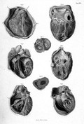
- Digital Images
- Online
The heart, it's cavities and valves. Fig. 1 - Anterior surface of Heart and Pericardial covering., Fig. 2 - Posterior surface of Heart, Auricles, and Ventricales, Fig. 3 - Internal cavities of Ventricals - anterior view, Fig. 4 - Anterior surface of Heart - interior of right auricle exposed, Fig 5 - Interior of right Auricle, Ventrical, and Pulmonary Artery, Fig. 6 - Exterior of left Ventricle and of Aorta, Fig. 7 - Transverse section of Auricles, Aorta, and Pulmonary Artery, immediately above the origins of these vessels, showing the auriculo-ventricular and arterial valves in action, Fig. 8 - Transverse section of Ventricles.
- Pictures
Post-mortem sketch of upper body showing venous gangrene in right arm of 74-year old woman with congestive heart failure and thrombosis causing several chronic occlusions. Watercolour by Barbara E. Nicholson, 1948.
Nicholson, BarbaraDate: 1948Reference: 32678iPart of: Barbara Nicholson medical illustration collection.